
This species forms dense carpets of straight to loose rope-like strands of dark green filaments (Image A). The uniseriate filaments are anchored by a rhizoidal basal cell augmented by both extramatrical (free of the main filament; Image B) and intramatrical (Images B & C) rhizoids descending from a few adjacent cells. Lower filaments are 27-31 µm wide, the cells squat to rectangular, 25-28 µm wide by 19-36 µm tall (Image D). Mid thallus filaments are 34-38 µm wide, the cells again slightly squat to rectangular, 28-32 µm wide by 24-50 µm tall (Image E). Upper thallus filaments are 52-100 µm wide, the cells more typically rectangular, 50-96 µm wide by 82-170 µm tall (Image F). Chloroplast are parietal and contain multiple pyrenoids (Image G). Zoidangia (gametangia?), 62-110 µm wide by 96-236 µm tall, are typically rectangular with a slight barrel appearance (Image H) and can be variable in length (Image I).
We have a single genetically verified record of this genetic group from high intertidal on rock in freshwater outflow at a sheltered location. It was tangled with considerable amounts of Urospora speciosa (Carmichael) Leblond ex Hamel (Image A) and a few filaments of Ulothrix implexa (Kützing) Kützing. This identification is at best tentative, but is at least superficially consistent with the detailed work of Lokhorst & Trask (1981).
This species is a larger version of Urospora sp. 2Nome, with which it is very closely allied in tufA sequence, these sister to Urospora wormskioldii (Mertens ex Hornemann) Rosenvinge. The latter two and the allied Urospora bangioides, as well as Urospora neglecta (Kornmann) Lokhorst & Trask, fail to join Urospora speciosa (Carmichael) Leblond ex Hamel and the type Urospora penicilliformis (Roth) Areschoug in our preliminary phylogenetic analyses. Taxonomic work is needed.
It is common for what otherwise looks like a unialgal carpet on rock, or mat of green filaments in an estuary, to be a chaotic mix of green species including, among others, various Blidingia spp., Percursaria percursa (C.Agardh) Rosenvinge, Rhizoclonium spp., Rosenvingiella polyrhiza (Rosenvinge) P.C.Silva, Urospora spp. and Ulothrix spp. Identification should be done carefully, as should attempting to match DNA sequences to individual species. Owing to the wonders of PCR, a clean sequence could be from any one of the species present in a collection and not necessarily the dominant one on which the microscopical identification was based. Consequently, I am not yet 100% certain that the genetic groups and morphospecies completely align for the various Urospora spp. presented on these pages. This is my best guess after consulting the pertinent literature (mostly Scagel (1966), Burrows (1991), Sears (2002), Brodie et al. (2007), Mathieson & Dawes (2017) and Gabrielson & Lindstrom (2018)). Considerable work remains for this genus in our flora.

Image A. Collection of this species forming loose rope-like strands (lower left) among abundant filaments of Urospora speciosa (Carmichael) Leblond ex Hamel (high intertidal on rock below freshwater outflow, culvert at head of Oak Bay, NB; GWS037969).

Image B. Attachment by a rhizoidal basal cell, as well as extramatrical and intramatrical rhizoids descending from adjacent cells (GWS037969; rehydrated from press).
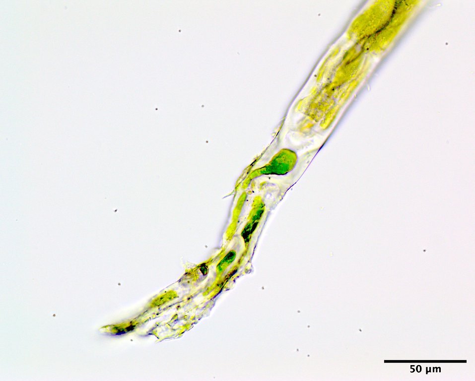
Image C. Holdfast dominated by intramatrical rhizoids (GWS037969; rehydrated from press).

Image D. Cells low on the filament (high intertidal in/on the steel culvert in brackish outflow, Culvert at head of Oak Bay, NB; GWS045939; Featured image above).

Image E. Cells mid filament (GWS045939).

Image F. Cells upper filament (GWS045939).
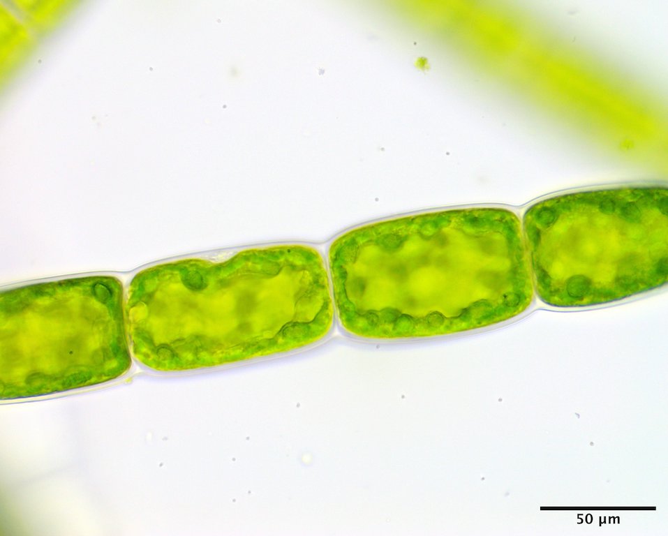
Image G. Chloroplast parietal with multiple pyrenoids in upper filament cells (GWS045939).

Image H. Zoidangia regularly rectangular with a slight swelling of the walls (GWS045939).
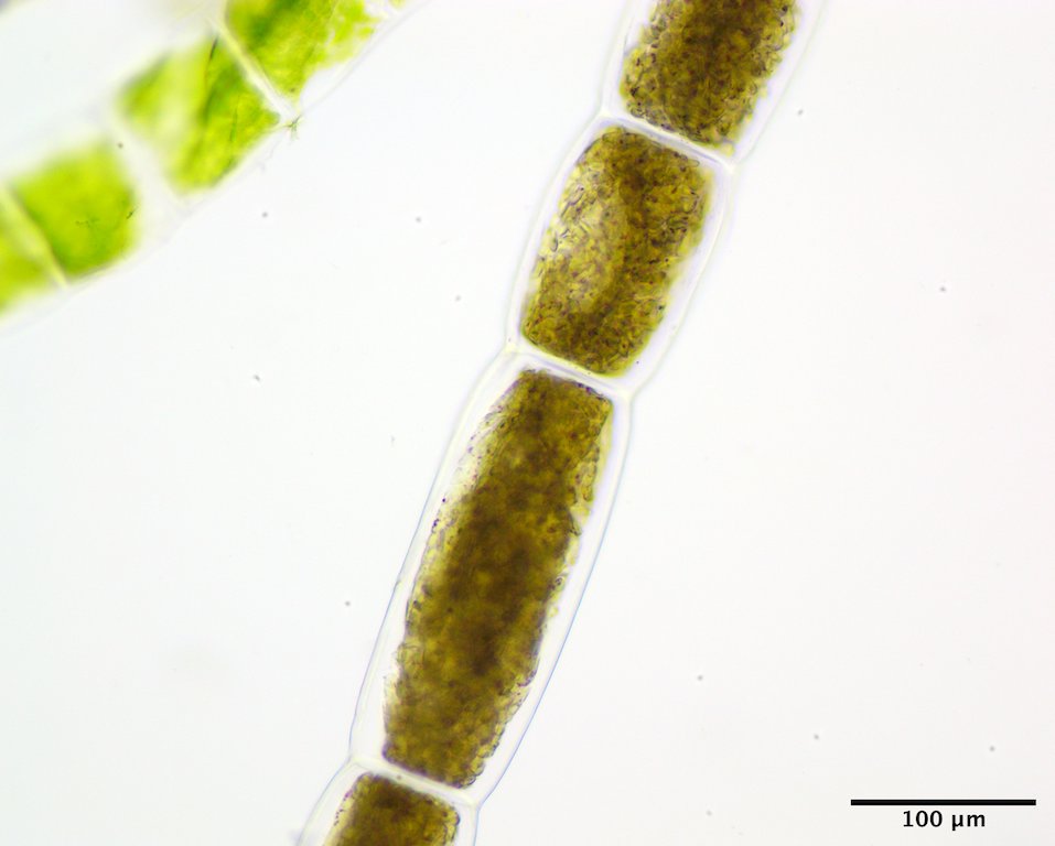
Image I. Zoidangia regularly rectangular with a slight swelling of the walls, but variable in length (GWS037969; rehydrated from press).
