Article

Adenophora triphylla var. japonica Inhibits Candida Biofilm Formation, Increases Susceptibility to Antifungal Agents and Reduces Infection

1
Graduate School of Biotechnology, Kyung Hee University, Seocheon, Giheung, Yongin 446-701, Gyeonggi-do, Korea
2
Department of Genetic Engineering, College of Life Science, Graduate School of Biotechnology, Kyung Hee University, Seocheon, Giheung, Yongin 446-701, Gyeonggi-do, Korea
*
Author to whom correspondence should be addressed.
Int. J. Mol. Sci. 2021, 22(22), 12523; https://doi.org/10.3390/ijms222212523
Submission received: 28 September 2021 / Revised: 31 October 2021 / Accepted: 18 November 2021 / Published: 21 November 2021
(This article belongs to the Section Molecular Microbiology)

Abstract

:
(1) Background: Candida is the most common cause of fungal infections worldwide, but due to the limited option of antifungal therapies, alternative strategies are required. (2) Methods: Adenophora triphylla var. japonica extract was used for the biofilm formation assay using RPMI1640. The combinatorial antifungal assay, the dimorphic transition assay, and the adherence assay were done to see the influence of inhibition of biofilm formation. qRT-PCR analysis were performed to check the gene expression. (3) Results: Adenophora triphylla var. japonica extract inhibited the Candida biofilm formation. Treatment of extract increased the antifungal susceptibility of miconazole from a 37% reduction in fungal growth to 99.05%, and also dose-dependently reduced the dimorphic transition of Candida and the attachment of Candida to HaCaT cells. The extract blocked the expression of hyphal-related genes, extracellular matrix genes, Ras1-cAMP-PKA pathway genes, Cph2-Tec1 pathway gene, and MAP kinase pathway gene. (4) Conclusions: In this study, the treatment of Adenophora triphylla var. japonica extract showed inhibition of fungal biofilm formation, activation of antifungal susceptibility, and reduction of infection. These results suggest that fungal biofilm formation is a good target for the development of antifungal adjuvants, and Adenophora triphylla var. japonica extract should be a good candidate for biofilm-associated fungal infections.

1. Introduction

Candida albicans is an opportunistic fungal pathogen that can cause systemic infections in individuals when mucosal barriers are disrupted, or the immune system is compromised [1]. Nosocomial C. albicans infection is often linked with the ability to form biofilms on mucosal surfaces and implanted medical devices [1,2,3].
Biofilm formation is a finely regulated process by multiple interconnected signaling pathways [4,5,6,7,8] and causes structured microbial communities to attach to surfaces [9,10]. The polysaccharide matrix indeed can act as a barrier for adherent cells by preventing the invasion of antifungal agents, conferring drug resistance. Adherent C. albicans cells living within the biofilm are up to 1000 times more resistant to common antifungal agents than planktonic cells, even in the absence of specific drug-resistance genes [11]. Thus, biofilms are a source of viable fungal cells that can potentially cause systemic infection with a mortality rate of 40–60% [12].
There have been many efforts to find novel natural compounds from medicinal plants to block biofilm-associated infections [13]. Medicinal plants are used in a variety of traditional remedies to treat cancer, infections, fever, asthma, and many other ailments, and generally have fewer side effects than over-the-counter medicines. Therefore, medicinal plants should be a new source of alternative therapies for the treatment of Candida infections [14,15].
Adenophora triphylla var. japonica (Korean name Zandae) has been used as an herbal medicine in Korea, China, and Japan to control lung diseases such as cough, sputum, asthma, and airway inflammatory diseases. Recently it was reported that extracts of A. triphylla var. japonica exhibited anti-inflammation, antidiabetic, antiparasitic, antitussive, and hepatoprotective effects [16,17,18].
In this study, A. triphylla var. japonica extract was tested for anti-biofilm-forming activity against several fungi including C. albicans, C. tropicalis, C. glabrata, and C. parapsilosis. Interestingly, the extract blocked biofilm formation, increased susceptibility to antibiotics, and reduced fungal infections.

2. Results

2.1. A. triphylla var. japonica Did Not Inhibit the Fungal Growth

Some antifungal agents inhibit fungal biofilm formation because they kill fungi and can indirectly reduce biofilm formation. A. triphylla var. japonica extract did not affect the growth of C. albicans after 24 h incubation. This result suggested that the extract’s inhibition of biofilm formation was not due to the reduced growth of Candida (Figure 1, Figure S1A–E)

2.2. A. triphylla var. japonica Extract Inhibited the Biofilm Formation of Candida

Biofilms are particularly important for fungi to survive and infect. A. triphylla var. japonica extract was used to test whether it blocks fungal biofilm formation. A. triphylla var. japonica extract dose-dependently inhibited Candida biofilm formation in all strains tested (Table 1) with an IC50 value of approximately 6.25 µg/mL against C. albicans (Figure 1). The IC50 value of the other strains was 6.25 μg/mL (C. parapsilosis var. parapsilosis and C. glabrata), 25 μg/mL (C. tropicalis var. tropicalis and C. tropicalis), and 50 μg/mL (C. parapsilosis). (Figure 2, Table 1).

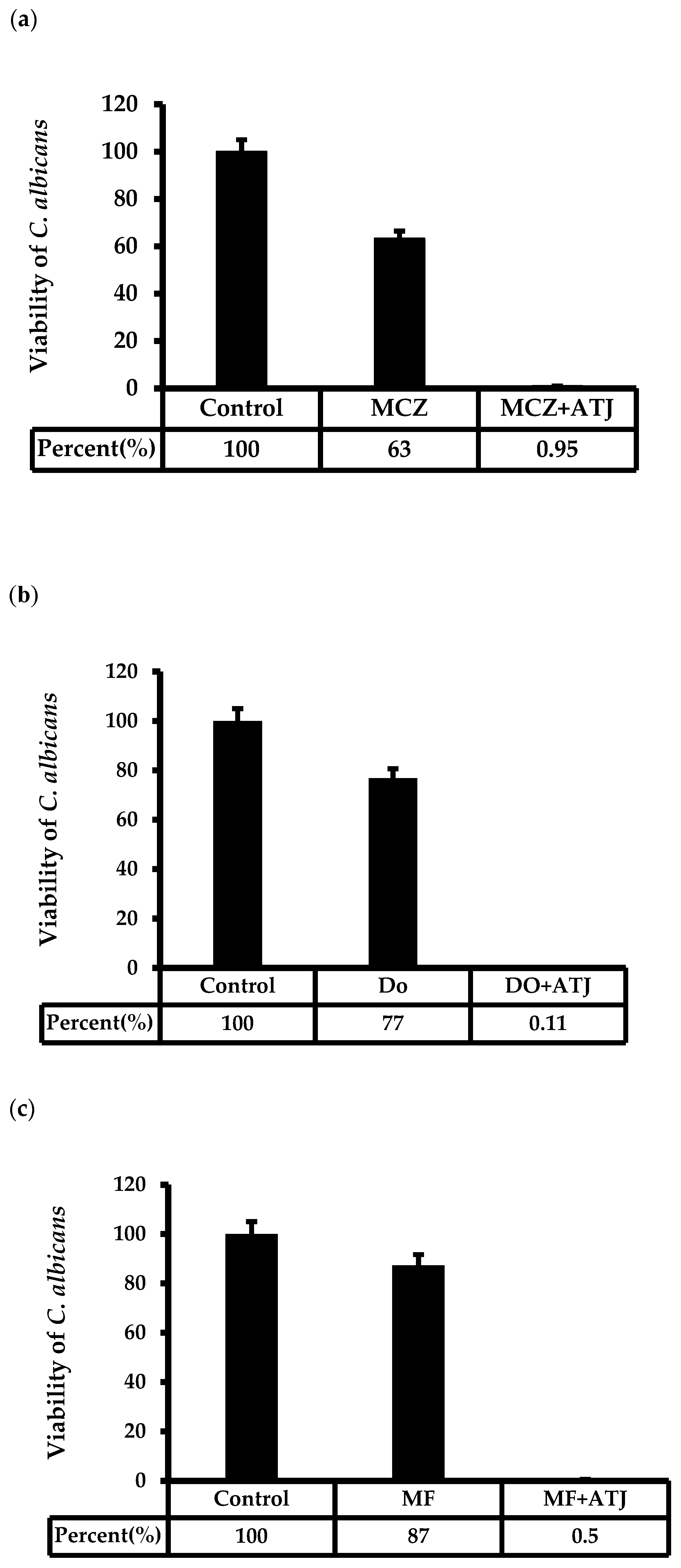
2.3. A. triphylla var. japonica Extract Increased the Susceptibility of Antifungal Agents against C. albicans

Biofilms are associated with antifungal activity; therefore, inhibition of biofilm formation is expected to increase antifungal efficacy. Treatment with 3.125 μg/mL of extract increased the susceptibility of miconazole from 38% inhibition to 99% inhibition of fungal growth. The extract also showed synergistic antifungal activity with magnoflorin (99% growth inhibition with 3.125 μg/mL extract compared with 13% by magnoflorin alone) and diosin (99% growth inhibition with 3.125 μg/mL extract compared with 23% by dioscin alone) (Figure 3a–c).

2.4. A. triphylla var. japonica Blocked the Dimorphic Transition from Yeast to Hyphae Form

The yeast-to-hyphae conversion is an important virulence factor for C. albicans. The formation of hyphae aids the subsequent invasive growth of C. albicans, infiltrating host tissues and causing systemic infection [6]. Extracts were tested to determine whether the dimorphic transition was affected and found that 1.56 μg/mL of extracts significantly inhibited dimorphic transition of 45% in RPMI1640 and 39% in 10% FBS YPD medium compare with untreated control (Figure 4).

2.5. A. triphylla var. japonica Extract Eeduced Fungal Adherence to the HaCaT Cells

Fungal biofilm formation for device-related infections is an important medical problem. A. triphylla var. japonica extract dose-dependently decreased the adhesion of Candida to human HaCaT cells by extract treatment (Figure 5).

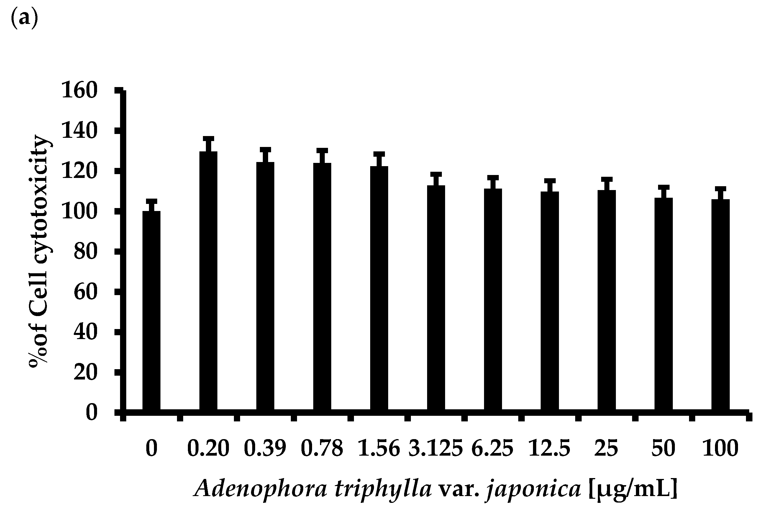
2.6. A. triphylla var. japonica Extract did Not Affect the Growth of Human Originated Cell

The cytotoxic effect of A. triphylla var. japonica extract on HaCaT cells and macrophages THP-1 was tested by MTT assay. A. triphylla var. japonica extract did not show any significant cytotoxic effect on both HaCaT and THP-1 cells (Figure 6a,b).

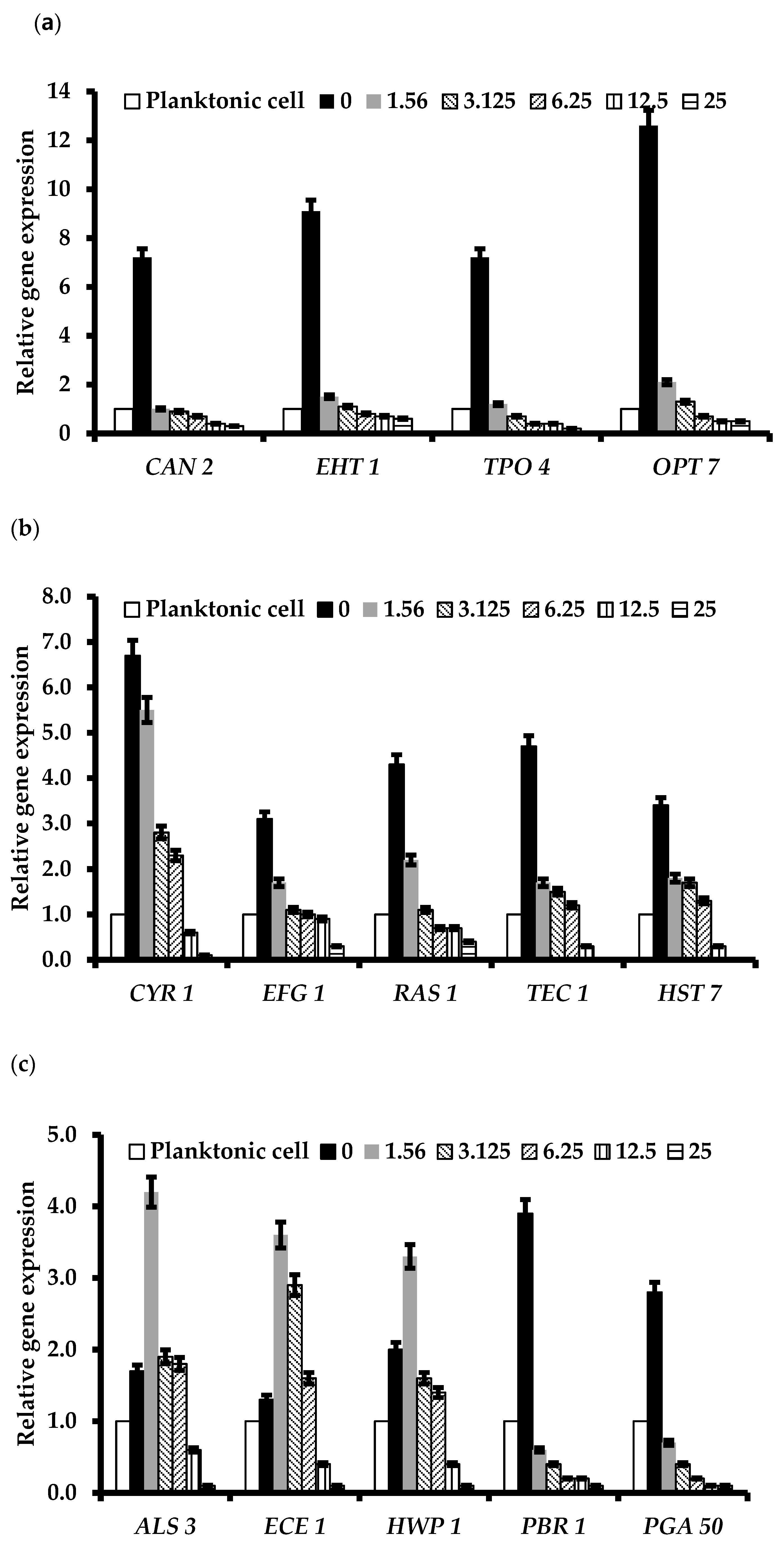
2.7. A. triphylla var. japonica Extract Inhibited the Expression of Biofilm Formation and Infection-Related Genes

To understand the molecular basis of the inhibition of biofilm formation and reduction of Candida infection, A. triphylla var. japonica extracts were tested for the expression of genes involved in biofilm formation, hyphae growth, and cell adhesion by qRT-PCR. The expressions of biofilm formation-related genes: [CAN2 (IC50 = 1.56 μg/mL), EHT1 (IC50 = 1.56 μg/mL), TPO4 (IC50 = 1.56 μg/mL), and OPT7 (IC50 = 1.56 μg/mL)]; Ras1-cAMP-PKA pathway related genes: [RAS1 (IC50 = 1.56 μg/mL), EFG1 (IC50 = 1.56 μg/mL), TEC1 (IC50 = 1.56 μg/mL), HST7 (IC50 = 1.56 μg/mL), and CYR1 (IC50 = 1.56 μg/mL)]; hyphal-specific genes: [ALS3 (IC50 = 12.5 μg/mL), ECE1 (IC50 = 12.5 μg/mL), and HWP1 (IC50 = 3.125 μg/mL)]; and extracellular matrix-related genes: [GSC1 (IC50 = 3.125 μg/mL), ADH5 (IC50 = 1.56 μg/mL), ZAP1 (IC50 = 3.125 μg/mL), and CSH1 (IC50 = 3.125 μg/mL)] were dose-dependently reduced by treatment of A. triphylla var. japonica extract (Figure 7a–d).

3. Discussion

Many fungi, including Candida, live inside the human body, but when Candida starts to grow out of control, it can cause an infection known as candidiasis. In fact, Candida is the most common cause of fungal infections in humans [1,2,3]. Antifungals can cause side effects and resistance, and there are several reports of resistance, including Candida auris, an emerging problematic organism [1,2,3]. Outbreak response is complicated by limited treatment options and inadequate disinfection strategies. Therefore, new approaches with novel targets for antifungal agents are needed.
In this study, A. triphylla var. japonica extract exhibited biofilm formation inhibitory activity against all Candida species tested (Figure 2, Table 1). The mechanism of Candida biofilm formation slightly differs depending on the species [5,6,7,8,9,10,11,12,13,14,15,16,17,18,19,20,21,22,23,24,25,26,27,28,29]. Comparisons are difficult because all Candida species have similar regulatory genes at each stage of biofilm formation. However, it can be emphasized that the ALS gene is involved in the adhesion process in three Candida species (C. albicans, C. parapsilosis, and C. tropicalis), and that this phenomenon is regulated by Epas in C. glabrata. In addition, some transcription factors described as being involved in C. albicans biofilm formation (BCR1, EFG1 and HWP1) are the same as those involved in C. parapsilosis biofilms. C. glabrata is a species that exhibits more contrast with respect to other Candida species, reflecting genetic distance [5]. A. triphylla var. japonica extract inhibits Candida biofilm formation, suggesting that there should be a common mechanism that is regulated by plant extract to influence the formation of biofilms among Candida species, but more studies are needed. In addition, A. triphylla var. japonica extract increased the antifungal activity of miconazole, magnoflorine, and dioscin (Figure 3), and decreased Candida infection (Figure 4 and Figure 5). These results showed that biofilm formation was associated with the susceptibility of antifungal agents and fungal infections.
According to gene expression analysis, the expression of genes related to biofilm formation, hyphae growth, and cell adhesion was significantly reduced by A. triphylla var. japonica extract treatment. Biofilm formation in Candida species is determined by various transcription factors, including BCR1, EFG1, TEC1, and NDT80, which act as components in multiple pathways, and their participation in Candida adhesion also suggests that although biofilm formation is initially tested, adhesion or infectious factors are also controlled by treatment with A. triphylla var. japonica extract. Further studies should be performed including the precise targeting of A. triphylla var. japonica extracts, effector components, and in vivo testing (Scheme 1).
In conclusion, A. triphylla var. japonica extracts inhibited C. albicans biofilm formation, increased antifungal activity of antifungal agents, and decreased fungal adhesion to host cells. Therefore, A. triphylla var. japonica extract should be a good candidate for biofilm-forming fungal infections.

4. Materials and Methods

4.1. Fungal Strains Used in This Study

The Candida strains used in the study are listed in Table 2. C. albicans KCTC 7965 and ATCC 10231 are the same strain. C. tropicalis KCTC 7212 was sourced from a patient with bronchomycosis. C. parapsilosis var. parapsilosis KACC 45480 and ATCC 22019 are the same strain. C. glabrata KCTC 7219 was sourced from faces. C. parapsilosis KACC 49573 was sourced from Canis familialis. All strains were stored in 20% glycerol at −70 °C and incubated on YPD plates [Peptone 20 g/L (BD Difco, Franklin Lakes, NJ, USA), yeast extract 10 g/L (BD Difco, Franklin Lakes, NJ, USA) and 2% glucose (w/v) (Daejung, Korea)] [27,28,29,30].

4.2. Extraction of A. triphylla var. japonica

The leaves of A. triphylla var. japonica were obtained from Jeju Island. Three-hundred grams of the sample was extracted using 3 L distilled water at 80 °C for 8 h, concentrated at 40 °C using a rotary evaporator, and then freeze-dried. For the experiment, 10 mg of plant extract powder was dissolved in 1 mL of dimethyl sulfoxide [14,15,16,17,18,19].

4.3. Growth Curve Assay

Fungal cultures were prepared using fresh YPD at 1 × 10⁶ cell/mL of C. albicans [23,24,25,26,27,28,29]. One hundred µg/mL of A. triphylla var. japonica extract was added and incubated at 37 °C. Growth was assessed by measuring OD600 using a microplate reader after indicated time [23,24,25]. Experiments were performed in triplicate.

4.4. Biofilm Formation Assay

A. triphylla var. japonica extracts were prepared in 96-well plate plates (SPL, Korea) with the concentration ranging from 6.25 to 100 μg/mL. Wells without test compound served as controls (DMSO concentration 0.1%). C. albicans suspension at 1 × 10⁶ CFU/mL was prepared in RPMI 1640 medium [24,25,26,27,28,29,30,31]. Then, 100 μL of the solution was inoculated into a 96-well flat-bottom plate and incubated at 37 °C for 24 h with shaking. Biofilm formed was washed with PBS to remove nonadherent cells, and then 100 μL of 1% aqueous crystal violet was applied for 30 min. Each well was washed three times with PBS and destained with 150 μL of 30% acetic acid for 15 min. The absorbance was measured at 595 nm with a microplate reader (Bio Tek Instruments, Korea). Experiments were performed in triplicate.

4.5. Antifungal Activity Assay

C. albicans were grown overnight in YPD diluted to 1 × 10⁶ cells/mL. Induction of biofilm formation was performed as described above. The broth microdilution method was applied to determine the MICs using microplates [32]. Serial twofold dilutions of A. triphylla var. japonica extracts were prepared in YPD broth. The MIC value was determined as the minimum compound concentration. Miconazole (3.125 µg/mL), magnoflorin (3.125 µg/mL), and dioscin (3.125 µg/mL) alone or with the indicated concentration of A. triphylla var. japonica extract were added to each well and incubated for 24 h at 37 °C [23,24,25,26,27]. Experiments were performed in triplicate.

4.6. Dimorphic Transition Assay

C. albicans were grown overnight in YPD medium. 1 × 10⁶ cells/mL of Candida with or without extract were incubated in YPD medium supplemented 10% fetal bovine serum medium or sodium bicarbonate-free RPMI medium with glutamine buffered with MOPS [3-(N-morpholino) propanesulfonic acid] to pH 7 recommended by the CLSI standard to induce dimorphic transition for 4 h at 37 °C. Quantification of inhibition of dimorphic transition was performed by counting the number of yeast and hyphal cells in the population as previously described [25,26,27,28,29,30,31]. At least 1000 cells were counted in duplicate for each well and all assays were repeated five times. An image of the cells was obtained using a microscope.

4.7. Candida Adherence Assay

Human epithelial keratinocytes HaCaT cells were maintained in DMEM supplemented with 10% fetal bovine serum at 37 °C in a humidified atmosphere with 5% CO2. HaCaT cells (0.5 × 10⁶ cells per well) were grown to confluence for 24 h in 24-well plates.
1 × 10⁶ cells/mL of C. albicans mixed with indicated concentration of A. triphylla var. japonica extract was incubated at 37 °C for 24 h. After the DMEM was drained, the plate was carefully washed three times with PBS to remove non-adherent cells. The number of adherent C. albicans cells was measured by scraping the cells with a sterile scraper. Serial dilutions were plated on YPD agar to determine viability. A microscope was used to obtain representative images of the results [26,27].

4.8. MTT Assay

Cytotoxicity of A. triphylla var. japonica extracts against HaCaT and THP-1 was tested by a slightly modified MTT assay. 1 × 10⁴ HaCaT and THP-1 in DMEM and RPMI1640 media, respectively, were added to each well containing the indicated concentration of extracts and incubated for 24 h. After discarding the media, MTT (3-(4,5-dimethyl-thiazol-2-yl)-2,5-diphenyltetrazolium bromide, Sigma, St. Louis, MO, USA) in PBS was added into each well, followed by incubation for 3 h at 37 °C. The medium was then removed, and cells were suspended in 100 μL DMSO for 10m. Cell viability was calculated as optical density (OD540) values measured using a microplate reader (BioTek Instruments, Korea) and reported as a percentage compare with vehicle control [25]. Experiments were performed in triplicate.

4.9. qRT-PCR Analysis

C. albicans were grown overnight in YPD and diluted to 1 × 10⁶ cells/mL. The indicated concentration of A. triphylla var. japonica extract was incubated in RPMI1640 with shaking at 37 °C for 24 h. Total RNA was isolated using TRIzol reagent (Life Technology, Thermo Fisher Scientific, Waltham, MA, USA) according to the manufacturer’s instructions, and cDNA was obtained by reverse transcriptase (NanoHelix, Korea) reaction using 1 μg of RNA. qRT-PCR was performed with 2 × SybrGreen qPCR Mater Mix (CellSafe, Korea). Transcript levels of genes were calculated using the formula 2-ΔΔCT. The primer sequences used are listed in Table 3. ACT1 served as an internal control [23,24,25,26,27,28,29,30,31,32].

4.10. Statistical Analysis

All experiments were performed at least three times and data were presented as the ± mean S.D. Graphs were generated using Microsoft Excel (Microsoft Corp., Redmond, WA, USA).

Supplementary Materials

The following are available online at https://www.mdpi.com/article/10.3390/ijms222212523/s1.

Author Contributions

Data curation, D.K.; Funding acquisition, K.-Y.K.; Investigation, D.K.; Methodology, D.K.; Supervision, K.-Y.K.; Writing—original draft, D.K.; Writing—review & editing, D.K. and K.-Y.K. All authors have read and agreed to the published version of the manuscript.

Funding

This research was funded by the GRRC program of Gyeonggi province [GRRC-kyunghee2020(B04)].

Institutional Review Board Statement

Not applicable.

Informed Consent Statement

Not applicable.

Data Availability Statement

The data presented in this study are available on request from the corresponding author.

Acknowledgments

This research was funded by the GRRC Program of Gyeonggi province [GRRC-KyungHee2020(B04)], Republic of Korea.

Conflicts of Interest

The authors declare no conflict of interest.

References

  1. Cavalheiro, M.; Teixeira, M.C. Candida Biofilms: Threats, Challenges, and Promising Strategies. Front. Med. 2018, 5, 28. [Google Scholar] [CrossRef] [PubMed] [Green Version]
  2. Chandra, J.; Kuhn, D.M.; Mukherjee, P.K.; Hoyer, L.L.; McCormick, T.; Ghannoum, M.A. Biofilm formation by the fungal pathogen Candida albicans: Development, architecture, and drug resistance. J. Bacteriol. 2001, 183, 5385–5394. [Google Scholar] [CrossRef] [PubMed] [Green Version]
  3. Hasan, F.; Xess, I.; Wang, X.; Jain, N.; Fries, B.C. Biofilm formation in clinical Candida isolates and its association with virulence. Microbes Infect. 2009, 11, 753–761. [Google Scholar] [CrossRef] [Green Version]
  4. Hogan, D.A.; Sundstrom, P. The Ras/cAMP/PKA signaling pathway and virulence in Candida albicans. Futur. Microbiol. 2009, 4, 1263–1270. [Google Scholar] [CrossRef] [PubMed]
  5. Araújo, D.; Henriques, M.; Silva, S. Portrait of Candida Species Biofilm Regulatory Network Genes. Trends Microbiol. 2016, 25, 62–75. [Google Scholar] [CrossRef] [PubMed] [Green Version]
  6. Biswas, S.; Van, D.P.; Datta, A. Environmental sensing and signal transduction pathways regulating morphopathogenic determinants of Candida albicans. Microbiol. Mol. Biol. Rev. 2007, 71, 348–376. [Google Scholar] [CrossRef] [Green Version]
  7. Kim, M.-J.; Kil, M.; Jung, J.-H.; Kim, J. Roles of Zinc-responsive transcription factor Csr1 in filamentous growth of the pathogenic Yeast Candida albicans. J. Microbiol. Biotechnol. 2008, 18, 242–247. [Google Scholar]
  8. Lane, S.; Zhou, S.; Pan, T.; Dai, Q.; Liu, H. The Basic Helix-Loop-Helix Transcription Factor Cph2 Regulates Hyphal Development in Candida albicans Partly via Tec1. Mol. Cell. Biol. 2001, 21, 6418–6428. [Google Scholar] [CrossRef] [Green Version]
  9. Ramage, G.; Saville, S.P.; Thomas, D.P.; López-Ribot, J.L. Candida Biofilms: An Update. Eukaryot. Cell 2005, 4, 633–638. [Google Scholar] [CrossRef] [Green Version]
  10. Douglas, L. Candida biofilms and their role in infection. Trends Microbiol. 2003, 11, 30–36. [Google Scholar] [CrossRef]
  11. Ramage, G.; VandeWalle, K.; Lã³Pez-Ribot, J.L.; Wickes, B.L. The filamentation pathway controlled by the Efg1 regulator protein is required for normal biofilm formation and development in Candida albicans. FEMS Microbiol. Lett. 2002, 214, 95–100. [Google Scholar] [CrossRef] [PubMed] [Green Version]
  12. Wenzel, R.P.; Gennings, C. Bloodstream Infections Due to Candida Species in the Intensive Care Unit: Identifying Especially High-Risk Patients to Determine Prevention Strategies. Clin. Infect. Dis. 2005, 41, S389–S393. [Google Scholar] [CrossRef] [PubMed]
  13. Al-Fattani, M.A.; Douglas, L.J. Biofilm matrix of Candida albicans and Candida tropicalis: Chemical composition and role in drug resistance. J. Med. Microbiol. 2006, 55, 999–1008. [Google Scholar] [CrossRef]
  14. Siti, N.H.M.A.; Zainuddin, A.M.; Sharifah, R.W.A.; Lee, S.C.; Azizul, A.M.; Nor, A.Y. Herbal processing and extraction technologies. Sep. Purif. Rev. 2016, 45, 305–320. [Google Scholar]
  15. Nagappan, R. Evaluation of aqueous and ethanol extract of bioactive medicinal plant, Cassia didymobotrya (Fresenius) Irwin & Barneby against immature stages of filarial vector, Culex quinquefasciatus Say (Diptera: Culicidae). Asian Pac. J. Trop. Biomed. 2012, 2, 707–711. [Google Scholar]
  16. Cota, B.B.; De Oliveira, D.B.C.; Borges, T.C.; Catto, A.C.; Serafim, C.V.; Rodrigues, A.R.A.; Kohlhoff, M.; Zani, C.L.; Andrade, A.A. Antifungal activity of extracts and purified saponins from the rhizomes of Chamaecostus cuspidatus against Candida and Trichophyton species. J. Appl. Microbiol. 2020, 130, 61–75. [Google Scholar] [CrossRef]
  17. Gorovoi, P.; Ponomarchuk, G.; Strigina, L. A chemotaxonomic study on russian far-eastern campanulaceae. Phytochemistry 1971, 10, 2419–2423. [Google Scholar] [CrossRef]
  18. Park, S.H. Ethyl Acetate Fraction of Adenophora triphylla var. japonica inhibits migration of lewis lung carcinoma cells by suppressing macrophage polarization toward an M2 phenotype. J. Pharmacopunct. 2019, 22, 253–259. [Google Scholar]
  19. Chun, J.; Kang, M.; Kim, Y.S. A triterpenoid saponin from Adenophora triphylla var. japonica suppresses the growth of human gastric cancer cells via regulation of apoptosis and autophagy. Tumor Biol. 2014, 35, 12021–12030. [Google Scholar] [CrossRef]
  20. Ovesna, Z.; Vachalkova, A.; Horvathova, K. Taraxasterol and beta-sitosterol: New naturally compounds with chemoprotective/chemopreventive effects. Neoplasma 2004, 51, 407–414. [Google Scholar]
  21. Na, M.; Kim, B.Y.; Osada, H.; Ahn, J.S. Inhibition of protein tyrosine phosphatase 1B by lupeol and lupenone isolated from Sorbus commixta. J. Enzym. Inhib. Med. Chem. 2009, 24, 1056–1059. [Google Scholar] [CrossRef]
  22. Gachet, M.S.; Kunert, O.; Kaiser, M. Antiparasitic compounds from Cupania cinerea with activities against Plasmodium falciparum and Trypanosoma brucei rhodesiense. J. Nat. Prod. 2011, 75, 559–566. [Google Scholar] [CrossRef]
  23. Cho, J.; Choi, H.; Lee, J.; Kim, M.-S.; Sohn, H.-Y.; Lee, D.G. The antifungal activity and membrane-disruptive action of dioscin extracted from Dioscorea nipponica. Biochim. Biophys. Acta Biomembr. 2013, 1828, 1153–1158. [Google Scholar] [CrossRef] [Green Version]
  24. Kim, J.; Bao, T.H.Q.; Shin, Y.-K.; Kim, K.-Y. Antifungal activity of magnoflorine against Candida strains. World J. Microbiol. Biotechnol. 2018, 34, 167. [Google Scholar] [CrossRef] [PubMed]
  25. Lingmei, S.; Kai, L.; Dayoung, W. Effects of magnolol and honokiol on adhesion, yeast-hyphal transition, and formation of biofilm by Candida albicans. PLoS ONE 2015, 10, e0117695. [Google Scholar]
  26. Khodavandi, A.; Alizadeh, F.; Harmal, N.S.; Sidik, S.M.; Othman, F.; Jahromi, M.A.F.; Sekawi, Z.; Ng, K.-P.; Chong, P.P. Comparison between efficacy of allicin and fluconazole against Candida albicans in vitro and in a systemic candidiasis mouse model. FEMS Microbiol. Lett. 2011, 315, 87–93. [Google Scholar] [CrossRef] [Green Version]
  27. Paola, M.; Roberta, F.; Cosmeri, R.; Tavanti, A.; Lupetti, A. Inhibition of Candida albicans biofilm formation by the synthetic Lactoferricin derived peptide hLF1-11. PLoS ONE 2016, 11, e0167470. [Google Scholar]
  28. Zhang, L.L.; Lin, H.; Liu, W.; Dai, B.; Yan, L.; Cao, Y.B.; Jiang, Y.-Y. Antifungal Activity of the ethanol Extract from Flos Rosae Chinensis with activity against Fluconazole-Resistant Clinical Candida. Evid. Based Complement. Altern. Med. 2017, 2017, 4780746. [Google Scholar]
  29. Bonifácio, B.V.; Vila, T.V.M.; Masiero, I.F.; Silva, P.B.D.; Silva, I.C.D.; Lopes, É.D.O.; dos Santos Ramos, M.A.; Perez de Sousa, L.; Vilegas, W.; Bauab, T.M.; et al. Antifungal activity of a hydroethanolic extract from Astronium urundeuva leaves against Candida albicans and Candida glabrata. Front. Microbiol. 2019, 10, 2642. [Google Scholar] [CrossRef] [Green Version]
  30. Gacser, A. Adhesins in Candida parapsilosis: Understudied players in virulence. Virulence 2016, 7, 65–67. [Google Scholar] [CrossRef] [PubMed]
  31. Taff, H.T.; Nett, J.E.; Zarnowski, R.; Ross, K.M.; Sanchez, H.; Cain, M.T.; Hamaker, J.; Mitchell, A.P.; Andes, D.R. A Candida biofilm -induced pathway for matrix glycan delivery: Implications for drug resistance. PLoS Pathog 2012, 8, e1002848. [Google Scholar] [CrossRef] [PubMed] [Green Version]
  32. Clinical and Laboratory Standards Institute. Methods for Dilution Antimicrobial Susceptibility Tests for Bacteria That Grow Aerobically; Approved Standards, Document M7-A5; Clinical and Laboratory Standards Institute (CLSI): Wayne, PA, USA, 2006. [Google Scholar]
Figure 1. A triphylla var. japonica extract did not inhibit the growth of C. albicans. C. albicans (1 × 10⁶ cells/mL) containing 100 μg/mL of A. triphylla var. japonica extract was incubated for 24 h at 30 °C. Experiments were performed in triplicate.
Ijms 22 12523 g001
Figure 2. A. triphylla var. japonica extracts inhibited biofilm formation of C. albicans pp. Biofilm formation of C. albicans were induced in RPMI1640 with indicated concentrations of A. triphylla var. japonica extract for 24 h at 37 °C. Experiments were performed in triplicate.
Ijms 22 12523 g002
Figure 3. A. triphylla var. japonica increased the susceptibility of antifungal agent. A. triphylla var. japonica extract (6.25 μg/mL) was treated to C. albicans with (a) miconazole (3.125 μg/mL), (b) dioscin (3.125 μg/mL), and (c) magnoflorin (3.125 μg/mL). Biofilm formation was induced for 24 h by growing of C. albicans in the RPMI1640. MCZ: Miconazole, MF: Magnoflorine, Do: Dioscin, ATJ: A. triphylla var. japonica extract. Experiments were performed in triplicate.
Ijms 22 12523 g003
Figure 4. Inhibition of C. albicans dimorphic transition by A. triphylla var. japonica extracts. (a) RPMI 1640 was used to induce C. albicans dimorphic transition and (b) images of C. albicans cells were obtained using a microscope. A: C. albicans without extract, B: C. albicans treated with 1.56 μg/mL of extract, C: C. albicans treated with 6.25 μg/mL of extract, D: C. albicans treated with 25 μg/mL of extract. (c) 10% FBS YPD medium was used to induce C. albicans dimorphic transition and (d) images of C. albicans cells were obtained using a microscope. A: C. albicans without extract, B: C. albicans treated with 1.56 μg/mL of extract, C: C. albicans treated with 6.25 μg/mL of extract, D: C. albicans treated with 25 μg/mL of extract. Experiments were performed in triplicate.
Ijms 22 12523 g004
Figure 5. A. triphylla var. japonica extract inhibited the adherence of C. albicans to HaCaT cells. C. albicans (1 × 10⁶ cells/mL) containing the indicated concentration of A. triphylla var. japonica extract was incubated for 24 h at 37 °C and the remaining Candida cells on HaCaT cells were counted. Experiments were performed in triplicate.
Ijms 22 12523 g005
Figure 6. Effects of A. triphylla var. japonica extract on the cell viability in HaCaT cells and THP-1 cells. (a) HaCaT cells and (b) THP-1 cells were treated with the indicated concentration of A. triphylla var. japonica extract for 24 h. The cell viability was evaluated by MTT assay. Experiments were performed in triplicate.
Ijms 22 12523 g006aIjms 22 12523 g006b
Figure 7. A. triphylla var. japonica extract inhibited the expression of genes involved in biofilm formation and toxicity of C. albicans. A. triphylla var. japonica extract reduced the expression of genes related to (a) biofilm formation, (b) Ras1-cAMP-Efg1 pathway, (c) hyphae-specific, and (d) extracellular matrix. Total RNA was extracted from C. albicans treated with the indicated concentration of A. triphylla var. japonica extract using an RNA extraction kit, converted to cDNA, and analyzed by qPCR using each primer. Experiments were performed in triplicate.
Ijms 22 12523 g007aIjms 22 12523 g007b
Scheme 1. Schematic diagrams for the anti-candida effect of A. triphylla var. japonica extract.
Ijms 22 12523 sch001
Table 1. A. triphylla var. japonica extracts inhibited biofilm formation of Candida spp. IC50 value.
StrainIC50 (Half Maximal Inhibitory Concentration)
C. albicans6.25 µg/mL
C. tropicalis var. tropicalis25 μg/mL
C. parapsilosis var. parapsilosis6.25 μg/mL
C. glabrata6.25 μg/mL
C. tropicalis25 μg/mL
C. parapsilosis50 μg/mL
Table 2. Strains used in this study.
StrainDescriptionSource
C. albicansKCTC 7965Purchased form KCTC (Korean Collection for Type of Cultures) or KACC (Korean Agriculture Culture Collection)
C. tropicalis var. tropicalisKCTC17762
C. parapsilosis var. parapsilosisKACC45480
C. glabrataKCTC7219
C. tropicalisKCTC7212
C. parapsilosisKACC49573
Table 3. Gene specific primers used for real-time RT-PCR.
GenesPrimer SequenceGene FunctionReferences
ACT1F: TAGGTTTGGAAGCTGCTGG
R: CCTGGGAACATGGTAGTAC
Control[8]
CAN2F: GCGGAATGGATATGCATGGG
R: CGGATTGCTCTTGGAGAAGC
Biofilm formationIn this study
EHT1F: TCGGAAAGCTTGGTGAAAGC
R: ATTTGGCCAAAGCAGGACTC
Biofilm formationIn this study
TPO4F: GCGGAATGGATATGCATGGG
R: CGGATTGCTCTTGGAGAAGC
Biofilm formationIn this study
OPT7F: TTGATCCCAGCTGCCAAATG
R: TGGCCCAAGTTCTTCGTATC
Biofilm formationIn this study
CYR1F: GTTTCCCCCACCACTCA
R: TTGCGGTAATGACACAACAG
Ras1-cAMP-Efg1 pathway[8]
EFG1F: TTGAGATGTTGCGGCAGGAT
R: ACTGGACAGACAGCAGGAC
Ras1-cAMP-Efg1 pathway[8]
HST7F: GCCAGTATGGTCGGAGGAT
R: ACATAGGCATCGTCTTCGTC
MAP kinases pathway[8]
RAS1F: GAGGTGGTGGTGTTGGTA
R: TCTTCTTGTCCAGCAGTATC
Ras1-cAMP-Efg1 pathway[8]
TEC1F: GCACTGGCTTCAAGCTCAAA
R: GCTGCTGCACCAAGTTCTG
Extracellular matrix[8]
ALS3F: GGTTATCGTCCATTTGTTG
R: TTCTGTATCCAGTCCATCT
Hyphal-specific genes[8]
ECE1F: ACAGTTTCCAGGACGCCAT
R: ATTGTTGCTCGTGTTGCCA
Hyphal-specific genes[8]
HWP1F: ACAGGTAGACGGTCAAGG
R: GGGTAATCATCACATGGTTC
Ras1-cAMP-Efg1 pathway[8]
PBR1F: TGTTGCTGCTGGTTCTGATG
R: GGTGGCAGATTTGGATTACC
Hyphal-specific genes[8]
PGA50F: ATTTCGAAGGGTGCAACTGC
R: AAGCACTGCAATGGGAGTTG
Hyphal-specific genesIn this study
ADH5F: ACCTGCAAGGGCTCATTCTG
R: CGGCTCTCAACTTCTCCATA
Extracellular matrix[8]
CSH1F: CGTGAGGACGAGAGAGAAT
R: CGAATGGACGACACAAAACA
Extracellular matrix[8]
GSC1F: CCCATTCTCTAGGCACGA
R: ATCAACAACCACTTGCTTCG
Extracellular matrix[8]
ZAP1F: ATCTGTCCAGTGTTGTTTGTA
R: AGGTCTCTTTGAAAGTTGTG
Extracellular matrix[8]
Publisher’s Note: MDPI stays neutral with regard to jurisdictional claims in published maps and institutional affiliations.

