Article

Metabolism of Mycosporine-Glutamicol in the Lichen Cladonia arbuscula subsp. squarrosa under Seasonal Changes and Elevated Exposure to UV-B or PAR Irradiation

1
Department of Plant Physiology and Development, Faculty of Biochemistry, Biophysics and Biotechnology, Jagiellonian University, Gronostajowa 7, 30-387 Krakow, Poland
2
Department of Microbiology, Institute of Quality Sciences and Product Management, Cracow University of Economics, Rakowicka 27, 31-510 Krakow, Poland
*
Author to whom correspondence should be addressed.
Metabolites 2022, 12(7), 632; https://doi.org/10.3390/metabo12070632
Submission received: 10 June 2022 / Revised: 29 June 2022 / Accepted: 4 July 2022 / Published: 10 July 2022
(This article belongs to the Special Issue Metabolism of Plant Secondary Metabolites under Stress Conditions)

Abstract

:
Cladonia arbuscula in its environmental niches is regularly affected by daily and annual variations in solar radiation. Mycosporine-glutamicol, Myc-Glu(OH), which it synthesizes, may act as a significant cellular UV-protector. Therefore, we studied this compound concentration in lichen thalli concerning seasonal changes and increased exposure to UV-B and photosynthetically active radiation (PAR) with/without simultaneous CO2 deprivation. Myc-Glu(OH) occurred year-round and exhibited a strong seasonality. The most crucial role in the control of its synthesis played UV-B radiation, although its high concentration was also found after PAR irradiation at 1000 µmol m−2 s−1. As PAR intensity increased to 2000 µmol m−2 s−1, the rate of Myc-Glu(OH) synthesis slowed down. In turn, under dark/PAR irradiation with simultaneous deprivation of CO2 in the atmosphere surrounding C. arbuscula and during darkness with continuous access to atmospheric CO2, its production was insignificant. Obtained data confirmed that Myc-Glu(OH) plays an important role in protecting C. arbuscula from UV damage and favours its adaptation to environmental stress in its natural habitat. They also suggest that its synthesis is a synergism of multiple factors. Consequently, further studies should focus on their evaluation and the identification of a lichen partner actively involved in Myc-Glu(OH) biogenesis.

1. Introduction

Lichen Cladonia arbuscula is permanently subjected in its natural habitat to strong solar irradiation including photosynthetically active radiation (PAR) and ultraviolet radiation (UV). The excessive doses of UV-B (280–315 nm) can directly induce conformational modifications in DNA via the formation of cyclobutane-pyrimidine dimers (CPDs) and 6-4 pyrimidine-pyrimidine (6-4-PP) photoproducts leading to genetic mutations, whereas UV-A (315–400 nm) can trigger the production of reactive oxygen species (ROS) that promote cellular damage of nucleic acids, proteins and lipids [1,2].
The key feature of many taxonomically different organisms to successful colonization of sunlit niches is to develop a variety of strategies to prevent or counteract the deleterious effects of UV irradiation. Besides the DNA reparative mechanisms, they show a synthesis of antioxidants, detoxifying enzymes, and/or UV-screening compounds such as mycosporine-like amino acids (MAAs) [3,4,5,6,7,8,9,10]. MAAs are low-molecular metabolites characterized by a cyclohexenone or cyclohexenimine chromophore conjugated with a nitrogen moiety and the capability of UV absorption in the harmful range from 309 to 362 nm [1,9,11,12,13].
Our previous research revealed in C. arbuscula thalli the presence of MAA compound, which was identified as mycosporine-glutamicol, Myc-Glu(OH) [14] (Figure 1). Based on the obtained results, we hypothesized that this compound may act as the significant cellular UV-protector and its concentration might be a function of irradiation dose. Therefore, in the present study, we have investigated the kinetics of its accumulation (a) throughout the whole calendar year to verify its possible seasonal variation in natural community and (b) under laboratory conditions simulating elevated exposure to UV-B or PAR irradiation, with or without simultaneous deprivation of CO2, to understand the mechanisms of this adaptive strategy.

2. Materials and Methods

2.1. Experimental Organism

The experimental organism Cladonia arbuscula (Wallr.) Flot subsp. squarrosa (Wallr.) Ruoss was harvested in April 2014 in Sobkow-Wolica (50°44′17.4″ N, 20°25′56.1″ E), Poland, and identified based on morphological characteristics with the help of standard taxonomic keys [15,16]. A voucher specimen has been kept for future references in the herbarium of the Department of Plant Physiology and Development, Jagiellonian University, Krakow, Poland.

2.2. Experimental Conditions

The naturally wetted C. arbuscula thalli (15 g fresh weight each) were placed in small beakers and then in glass vessels ¼ filled with water. Immediately afterwards lichen-forming fungi were subjected to the effect of a specified factor:
  • PAR (λ = 400–700 nm) at the intensity of 1000, 1500, and 2000 µmol m−2 s−1 (two 1000 W Tungsram lamps, The Netherlands, 12 h light: 12 h dark) or darkness (24 h) with continuous access to atmospheric CO2 levels;
  • UV-B radiation (λ = 290–315 nm, λmax = 310 nm) of 5 µmol m−2 s−1 (two fluorescence tubes, Philips TL 40W/12, Germany, screened with a 0.13 mm thick cellulose acetate filter paper to remove all radiation below 290 nm);
  • PAR of 1000 µmol m−2 s−1 or darkness with simultaneous deprivation of CO2 in the atmosphere surrounding lichen-forming fungus, which was achieved by replacing water in a glass vessel with a 5% Ba(OH)2 solution.
Under the above conditions, C. arbuscula was cultivated over 49 days in the growth chamber at 21 ± 1 °C and humidity of 80%. Irradiation intensity was measured inside the vessels using a Photometer-Radiometer (HD 2102.1/2102.2, Delta OHM, Selvazzano Dentro, Italy) with LP471UVB and LP471PAR probes. Samples for analysis were collected at 0, 7th, 14th, 28th, and 49th day of treatment. The material was divided into the upper (tip) and lower (stem) parts of podetia.

2.3. Kinetics of the Changes in Myc-Glu(OH) Concentration

To evaluate the concentration of Myc-Glu(OH) in C. arbuscula, during the changing seasons, its thallus was collected cyclically 4 times a month at weekly intervals in 2014.

2.4. Sample Preparation

All samples of the lichen-forming fungus were air-dried, crushed, weighed in equal portions, and immersed in 25% methanol (MeOH) (v/v) for 3 h at 45 ± 1 °C according to the procedure described by Tartarotti and Sommaruga [17]. The supernatants were filtrated through WhatmanTM glass filters GF/C (USA) ∅ 0.45 μm, centrifuged (10,000× g, 5 min), and evaporated to dryness under reduced pressure at 21 ± 1 °C. Pellets were re-suspended in 100% MeOH, and aliquots were re-centrifuged and re-evaporated. The dry residues were re-dissolved in Milli-Q water followed by the addition of amounts of chloroform with gentle vortexing. After centrifugation, the uppermost water phases were filtered through the Durapore syringe filters (Durapore, Germany) ∅ 0.22 μm and kept at −20 ± 1 °C for high-pressure liquid chromatography (HPLC) analysis.

2.5. Analytical Determination

Chromatographic analyses were conducted using a Waters (Waters, Milford, MA, USA) apparatus consisting of a 600E gradient pump, 717 plus autosampler, 996 photodiode detector, Millenium32 SS Software, and a Jetstream 2 plus column oven. Qualitative and quantitative identification of Myc-Glu(OH) was performed on an analytical column Waters Atlantis® dC18 (3.9 × 100 mm; 3 μm) with guard column C18 thermostated at 35 ± 1 °C by using a mobile phase consisting of water/acetonitrile (ACN); both were acidified with 0.05% trifluoroacetic acid (TFA, v:v). A linear gradient was set from 100% to 77% water in 6 min at a flow rate of 1 mL min−1. The Myc-Glu(OH) was identified by comparing the UV spectra determined for prepared standard and quantified by absorbance at λmax = 309 nm using the calibration curve.

2.6. Reagents and Chemicals

All the chemicals were analytical or HPLC grade and were purchased from Sigma-Aldrich (USA). Milli-Q water was obtained using an Elga (Elga LabWater, High Wycombe, UK) Maxima LS Ultra-Pure Water Purifier.

2.7. Statistical Analysis

The results were presented as means ± standard deviation (S.D.) of five independent replicates. Data were analysed using analysis of variance (ANOVA) in Statistica 10 software (StatSoft, Tulsa, OK, USA). Differences were considered significant at p < 0.05 [18].

3. Results and Discussion

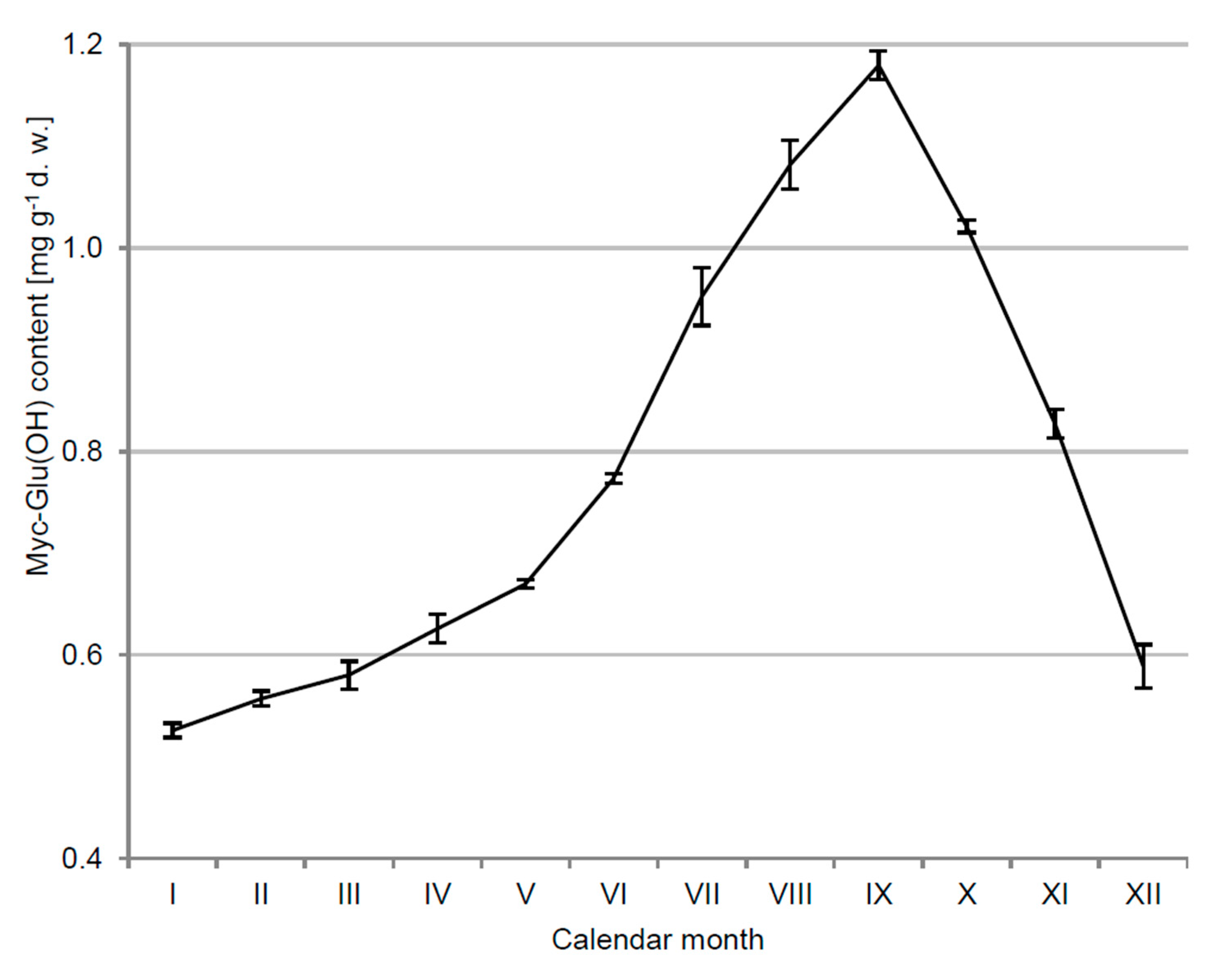
3.1. Kinetics of Myc-Glu(OH) Concentration in C. arbuscula Thalli

C. arbuscula in its ecological niches is regularly affected by daily and annual variations in solar radiation. For lichen-forming fungi, numerous studies show a strong correlation between the environmental factors and the intensity of secondary metabolites production [19]. This connection also refers to the biosynthesis of Myc-Glu(OH). Its concentration in C. arbuscula thalli exhibited a clear seasonality, ranging from 0.526 ± 0.007 mg g−1 of dry weight (d.w.) in January to 1.179 ± 0.014 mg g−1 d.w. in September 2014 (>2-fold increase) (Figure 2). The elevational trend in Myc-Glu(OH) accumulation over the summer was correlated with the greater intensity of solar energy reaching the Earth’s surface and clearly confirmed its relevance in acclimatization of the lichen-forming fungus to such conditions. In turn, a decrease in the concentration of this highly polar and water-soluble compound in October was probably related to its leaching out from the thallus by rainwater. Previously, potential seasonal fluctuations in the quantitative and qualitative composition of MAAs have been verified only for freshwater and marine species [20,21,22,23,24,25,26]. However, they did not demonstrate one common pattern for all tested organisms. A similar annual variation to the Myc-Glu(OH) occurrence was reported for MAAs in the phytoplankton cells and copepod Cyclops abyssorum tatricus body from a transparent alpine lake [24]. Both organisms showed, respectively, a 3.6 and 3-fold higher level of MAAs synthesis in the summer compared to winter months. The MAAs concentration in phytoplankton collected from the English Channel was also closely correlated with seasonal changes in solar radiation intensity [22], whereas such a pattern was not observed for the Antarctic sea urchin Sterechinus neumayeri [20]. Comparable annual variability to the Myc-Glu(OH) accumulation was also revealed for other photoprotective compounds. The intensity of melanin production by the subarctic Daphnia spp. significantly increased after the ice-break [27], and the concentration of parietin in the lichen Xanthoria parietina exhibited a rapid spring growth and an autumn decline [28].
Generally, MAAs concentration does not exceed 1% of dry weight [1]. Similar values to the Myc-Glu(OH) occurrence in C. arbuscula thalli were determined for mycosporine-glycine in the lichen Lichina pygmae; the concentration of this compound was equal to 1.11 ± 0.23 mg g−1 d.w. [29]. A significantly higher mycosporine-glycine concentration, from 0.1 to 0.8% of d.w., characterized lichens belonging to the Collema, Gonohymenia, and Peltula genera [30].

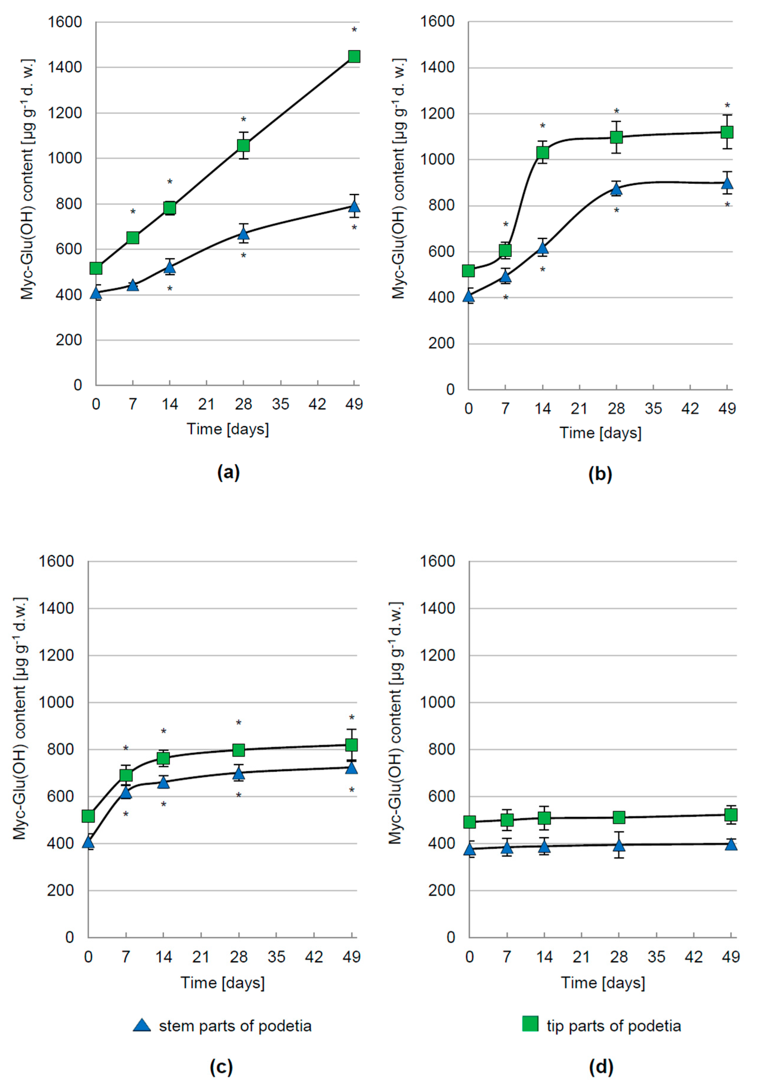
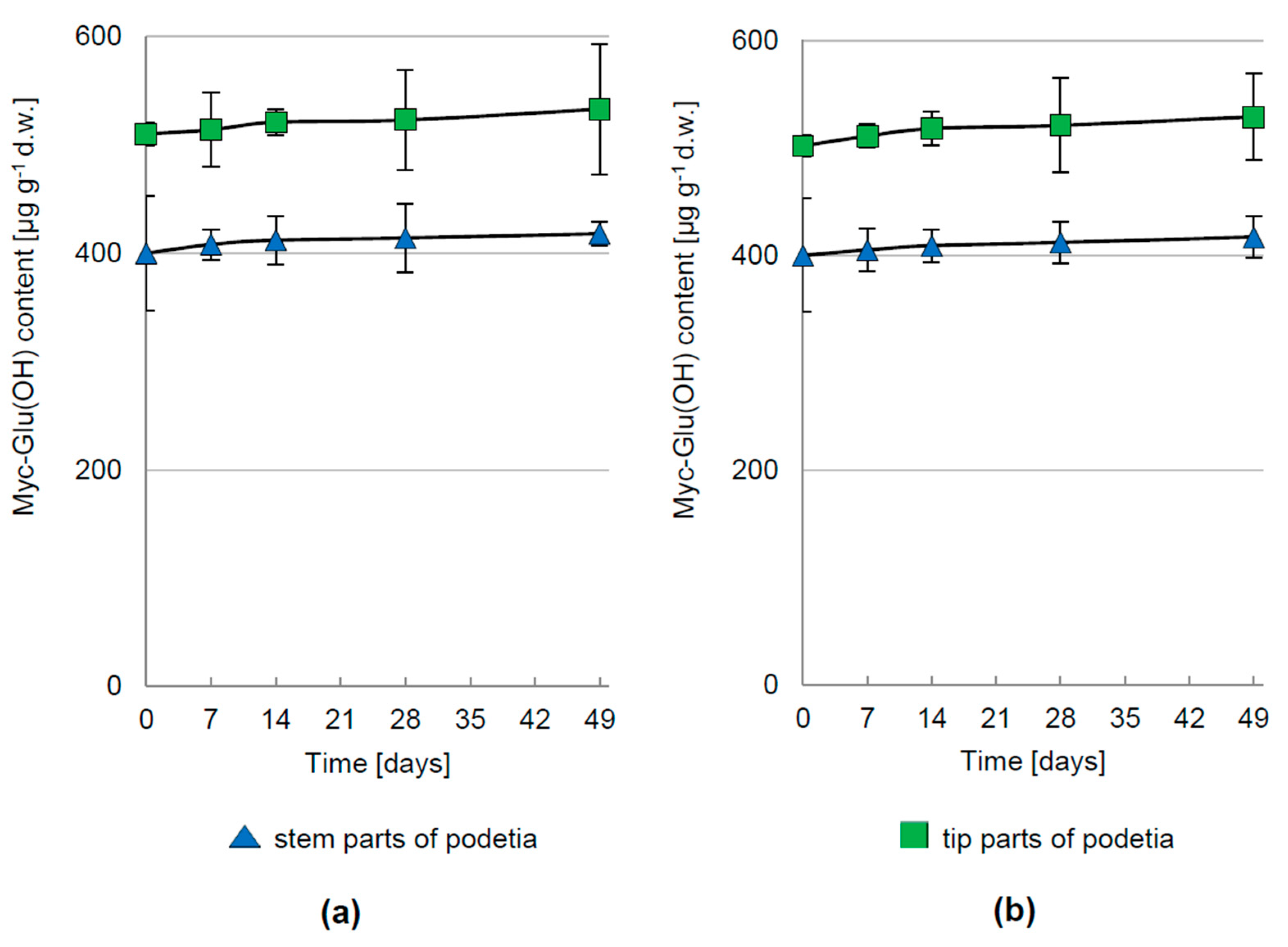
3.2. Regulation of Myc-Glu(OH) Accumulation

In vivo experiments showed that the production of Myc-Glu(OH) is controlled not only by different intensity but also the spectral composition of solar rays. PAR was one of the important factors determining the synthesis level of Myc-Glu(OH) in C. arbuscula thalli, but the kinetics of this process depended on the applied intensity. The greatest increase in its concentration by 180% (1448 µg g−1 d.w.) and 93% (791 µg g−1 d.w.) in the tip and stem parts of podetia, respectively, was determined after 49 days of treatment with 1000 µmol m−2 s−1 (Figure 3a). Along with the elevation of PAR radiation up to 1500 µmol m−2 s−1, the rate of Myc-Glu(OH) production slowed down and at the end of the experiment its accumulation enhanced by 117% (1120 µg g−1 d.w.) in the tip and 120% (900 µg g−1 d.w.) in the stem parts of podetia compared to their initial values (Figure 3b). In turn, the highest tested PAR dose of 2000 µmol m−2 s−1 stimulated the Myc-Glu(OH) production only at the beginning of the cultivation, followed by the stabilization period (Figure 3c). Finally, its concentration was equal to 820 µg g−1 d.w. in the tip (an increase of 77%) and 724 µg g−1 d.w. in the stem parts of podetia (an increase of 59%). The lower level of metabolite synthesis under higher light intensities may be the effect of photosynthetic apparatus damage.
Similar hypersensitivity was documented in hydrated thalli of several lichen species [31]. The elevated production of MAAs and changes in their composition under PAR irradiation was also demonstrated for the dinoflagellate Alexandrium excavatum moving from low (20 μE m−2 s−1) to 10 times greater light intensities [32]. In turn, a 14-fold higher concentration of MAAs was reported for the dinoflagellate Gymnodinium sanguineum cultured in the cold white fluorescent light of approx. 350 μmol m−2 s−1 compared to those treated with approx. 70 μmol m−2 s−1 [33]. MAAs accumulation was also induced by different spectral compositions (white, blue, green, yellow, and red light). Korbee et al. described the beneficial role of blue light on porphyra-334, palythine, and asterine-330 synthesis in the red alga Porphyra leucosticta, whereas white, green, yellow, and red light favoured the production of shinorine [34].
Despite the unquestionable importance of PAR in the control of the Myc-Glu(OH) synthesis in C. arbuscula thalli, the most crucial role in this process was played by UV-B radiation. Under 7-weeks exposition to 5 μmol m−2 s−1 the concentration of this metabolite was significantly elevated by 624% (3758 μg g−1 d.w.) and 457% (2282 μg g−1 d.w.) in the tip and stem parts of the podetia, respectively (Figure 4). The positive influence of UV-B on MAAs accumulation was also observed in the polar algae Phaeocystis antarctica [35], dinoflagellates Alexandrium tamarense and Heterocapsa triquetra [36], and nitrogen-fixing cyanobacteria [6]. Compared to other wavelengths, UV-B exerted a more pronounced influence on MAAs synthesis in cyanobacteria [37]. Arthrospira sp. CU2556 and Gloeocapsa sp. treated simultaneously with PAR + UV-A + UV-B exhibited an effective increase in the production of mycosporine-glycine [38], shinorine, and M-307 [39], rather than irradiated with PAR or PAR + UV-A. The induction of palythine and mycosporine-glycine synthesis by the marine red alga Devaleraea ramentacea was also revealed after exposure to PAR in combination with UV-A and UV-B [40]. In contrast, the concentration of usnic acid (depsidone) and atranorin (depside), synthesized in addition to Myc-Glu(OH) by studied lichen-forming fungus, was affected mainly by UV-A [41]. Similar wavelength ranges stimulated the production of MAAs by Arctic diatoms [35,42,43]. However, opposite to the above examples, for some species such correlations have not been recorded at all [44].
De novo MAAs synthesis is a highly energetic and costly process; nevertheless, these costs are disproportionate to UV-induced damage [23,45]. Their presence in a cell can effectively prevent 3 out of 10 photons from hitting cytoplasmic targets [46]. Thus, an increase in the production rate of MAAs, including Myc-Glu(OH), due to excessive exposure to high doses of UV effectively reduces metabolic costs until the ecological benefits of their presence will be achieved [47].
Distribution of Myc-Glu(OH) within the C. arbuscula thalli irradiated with PAR or UV-B was not homogeneous. The difference in its concentration probably reflects (1) various metabolic activity of the enzymes controlling its synthesis in the upper, younger parts and in the lower, older parts of podetia [48]; (2) evolutionary adaptation of this lichen-forming fungus to UV protection of upper parts containing reproductive structures such as pycnidia; (3) a different degree of the exposure of the particular thalli parts to UV. The tip parts of podetia form branches that naturally shade the lower parts [49]; therefore, the enhanced synthesis of Myc-Glu(OH) by the latter may not be necessary. Similar dependence, as for the tested Myc-Glu(OH), was demonstrated for usnic acid in Flavocetraria nivalis [50] and phenolic compounds that effectively block the amount of UV-B penetrating into Cladonia mitis thalli [49].
Under dark or PAR irradiation (1000 μmol m−2 s−1) conditions with simultaneous deprivation of CO2 in the atmosphere surrounding C. arbuscula, the Myc-Glu(OH) concentration remained constant over the whole cultivation period (Figure 5a,b). Its comparatively insignificant production was also demonstrated during darkness with continuous access to atmospheric CO2 levels (Figure 3d).
Therefore, it is suggested that the synthesis of the analysed metabolite is dependent on the availability of light and CO2. Based on these results, we assume that the carbon compounds to Myc-Glu(OH) production are formed during the green algal photosynthesis rather than the fungal dark carboxylation. On the other hand, Myc-Glu(OH) possesses a carbonyl group characteristic for fungal mycosporines, which may suggest a significant contribution of mycobiont in its biogenesis. This hypothesis is also supported by the fact that Myc-Glu(OH) has been detected so far in the microcolonial fungi and sclerotia of the deuteromycete Botrytis cinerea [51,52,53,54]. Furthermore, the literature data clearly show that many of the UV-absorbing compounds, such as polyphenolics, anthraquinone pigments, or usnic acid are produced by mycobionts [30]. Nevertheless, both lichen partners may be involved in the biogenesis of Myc-Glu(OH); especially because the fungi and green algae, as independent organisms, are known for MAAs synthesis [55]. Only a few studies have shown that the production of MAAs has been clearly defined and attributed to one particular component. Torres et al. determined that the collemine A is produced by mycobiont of the Collema cristatum [56]. In addition, Roullier et al. revealed that cephalodia of the Stereocaulon philippinense, composed of cyanobacterial cells, contained MAA with λmax at 340 nm instead of the compound present in the whole thalli with λmax = 310 nm [57]. They assumed that one partner could synthesize MAA with λmax = 310 nm, and the other structurally modified it to the form with λmax = 340 nm. Although the parallel, uncorrelated and independent biogenesis of those two compounds in different parts of the thalli cannot be excluded. Some cyanobacteria, due to the ability to atmospheric nitrogen fixation, appear to be involved in the production of MAAs in cyanolichens.

4. Conclusions

The annual pattern of Myc-Glu(OH) content exhibited a clear seasonality with a more than 2-fold increase in concentration during elevated exposure to solar radiation in late summer. Its biosynthesis is regulated by UV-B and PAR, the effect of the latter being markedly weaker. These findings are consistent with the idea that this compound provides adaptation of C. arbuscula to relatively high radiation levels and irradiance changes in the natural habitat. Nevertheless, the enhanced biosynthesis of Myc-Glu(OH) is unquestionably a synergistic effect of many factors rather than individual PAR or UV-B influence as demonstrated by experiments with/without the presence of CO2 in the atmosphere surrounding lichen. Consequently, further research should focus on understanding these factors. Given that the accumulation of Myc-Glu(OH) in the C. arbuscula thalli is a benefit of this symbiosis, it would be useful to concentrate future studies on the identification of lichen partners actively involved in its biogenesis.

Author Contributions

Conceptualization, E.C.-S. and J.B.; methodology, E.C.-S., J.B., and B.B.; formal analysis, E.C.-S.; investigation, E.C.-S. and K.D.; data curation, E.C.-S.; writing—original draft preparation, E.C.-S.; writing—review and editing, J.B., B.B., and K.D.; visualization, E.C.-S.; supervision, J.B. and B.B.; funding acquisition, E.C.-S. All authors have read and agreed to the published version of the manuscript.

Funding

This research was funded by the National Science Centre (NCN), Poland, grant number 2015/17/N/NZ8/01575. Ewelina Chrapusta-Srebrny also received financial resources for the preparation of a doctoral dissertation from the NCN as part of the scholarship funding, grant number 2015/16/T/NZ8/00153.

Institutional Review Board Statement

Not applicable.

Informed Consent Statement

Not applicable.

Data Availability Statement

The data presented in this study are available on request from the corresponding author. The data are not publicly available due to the fact that they were provided in the manuscript.

Acknowledgments

We are grateful to Zbigniew Lechowski for his thoughtful comments, valuable advice, and assistance. We would also like to thank members of the Department of Plant Physiology and Development for reading the draft version of manuscript and two anonymous reviewers for greatly improving the paper. Faculty of Biochemistry, Biophysics and Biotechnology was a partner of the Leading National Research Center (KNOW) supported by the Ministry of Science and Higher Education.

Conflicts of Interest

The authors declare no conflict of interest.

References

  1. Karentz, D. Chemical defenses of marine organisms against solar radiation exposure: UV-absorbing mycosporine-like amino acids and scytonemin. In Marine Chemical Ecology; McClintock, J.B., Baker, J., Eds.; CRC Press, Inc.: Boca Raton, FL, USA, 2001; pp. 481–520. ISBN 978-0-8493-9064-7. [Google Scholar]
  2. Rastogi, R.P.; Sinha, R.P.; Singh, S.P.; Häder, D.-P. Photoprotective compounds from marine organisms. J. Ind. Microbiol. 2010, 37, 537–558. [Google Scholar] [CrossRef] [PubMed]
  3. Sinha, R.P.; Häder, D.-P. UV-induced DNA damage and repair: A review. Photochem. Photobiol. Sci. 2002, 1, 225–236. [Google Scholar] [CrossRef] [PubMed]
  4. Häder, D.-P.; Sinha, R.P. Solar ultraviolet radiation-induced DNA damage in aquatic organisms: Potential environmental impact. Mutat. Res. 2005, 571, 221–233. [Google Scholar] [CrossRef]
  5. Mittler, R.; Tel-Or, E. Oxidative stress responses in the unicellular cyanobacterium Synechococcus PCC 7942. Free Radic. Res. Commun. 1991, 12-13 Pt 2, 845–850. [Google Scholar] [CrossRef]
  6. Sinha, R.P.; Klisch, M.; Walter Helbling, E.; Häder, D.P. Induction of mycosporine-like amino acids (MAAs) in cyanobacteria by solar ultraviolet-B radiation. J. Photochem. Photobiol. B Biol. 2001, 60, 129–135. [Google Scholar] [CrossRef]
  7. Sinha, R.P.; Klisch, M.; Gröniger, A.; Häder, D.-P. Ultraviolet-absorbing/screening substances in cyanobacteria, phytoplankton and macroalgae. J. Photochem. Photobiol. B Biol. 1998, 47, 83–94. [Google Scholar] [CrossRef]
  8. Oren, A.; Gunde-Cimerman, N. Mycosporines and mycosporine-like amino acids: UV protectants or multipurpose secondary metabolites? FEMS Microbiol. Lett. 2007, 269, 1–10. [Google Scholar] [CrossRef]
  9. Chrapusta, E.; Kaminski, A.; Duchnik, K.; Bober, B.; Adamski, M.; Bialczyk, J. Mycosporine-Like Amino Acids: Potential Health and Beauty Ingredients. Mar. Drugs 2017, 15, 326. [Google Scholar] [CrossRef] [Green Version]
  10. Shang, J.-L.; Zhang, Z.-C.; Yin, X.-Y.; Chen, M.; Hao, F.-H.; Wang, K.; Feng, J.-L.; Xu, H.-F.; Yin, Y.-C.; Tang, H.-R.; et al. UV-B induced biosynthesis of a novel sunscreen compound in solar radiation and desiccation tolerant cyanobacteria. Environ. Microbiol. 2018, 20, 200–213. [Google Scholar] [CrossRef]
  11. Rastogi, R.P.; Singh, S.P.; Häder, D.-P.; Sinha, R.P. Mycosporine-like amino acids profile and their activity under PAR and UVR in a hot-spring cyanobacterium Scytonema sp. HKAR-3. Aust. J. Bot. 2010, 58, 286–293. [Google Scholar] [CrossRef]
  12. Shick, J.M.; Dunlap, W.C. Mycosporine-like amino acids and related gadusols: Biosynthesis, accumulation, and UV-protective functions in aquatic organisms. Annu. Rev. Physiol. 2002, 64, 223–262. [Google Scholar] [CrossRef] [PubMed] [Green Version]
  13. Singh, S.P.; Kumari, S.; Rastogi, R.P.; Singh, K.L.; Sinha, R.P. Mycosporine-like amino acids (MAAs): Chemical structure, biosynthesis and significance as UV-absorbing/screening compounds. Indian J. Exp. Biol. 2008, 46, 7–17. [Google Scholar] [PubMed]
  14. Chrapusta-Srebrny, E.; Białczyk, J.; Duchnik, K. Kinetics of mycosporine-glutamicol biosynthesis in vivo in Cladonia arbuscula (Wallr.) Flot subsp. squarrosa (Wallr.) Ruoss thalli. In Abstracts of Lectures and Posters of the 58th PBS Congress Botany without Borders—58th Congress of the Polish Botanical Society; Frey, L., Ed.; Polish Botanical Society: Kraków, Poland, 2019; p. 173. ISBN 978-83-954123-0-1. [Google Scholar]
  15. Nash, T.H., III (Ed.) Lichen Biology, 2nd ed.; Cambridge University Press: Cambridge, UK, 2008. [Google Scholar]
  16. Orange, A.; James, P.W.; White, F.J. Microchemical Methods for the Identification of Lichens; British Lichen Society: London, UK, 2001. [Google Scholar]
  17. Tartarotti, B.; Sommaruga, R. The effect of different methanol concentrations and temperatures on the extraction of mycosporine-like amino acids (MAAs) in algae and zooplankton. Arch. fur Hydrobiol. 2002, 1544, 691–703. [Google Scholar] [CrossRef]
  18. Stanisz, A. Przystępny kurs statystyki z zastosowaniem Statistica PL na przykładach z medycyny; StatSoft Polska: Kraków, Poland, 2006; ISBN 83-88724-18-5. [Google Scholar]
  19. Ranković, B.; Kosanić, M. Lichens as a Potential Source of Bioactive Secondary Metabolites. In Lichen Secondary Metabolites: Bioactive Properties and Pharmaceutical Potential; Ranković, B., Ed.; Springer International Publishing: Cham, Switzerland, 2015; pp. 1–26. ISBN 978-3-319-13374-4. [Google Scholar]
  20. Karentz, D.; Dunlap, W.C.; Bosch, I. Temporal and spatial occurrence of UV-absorbing mycosporine-like amino acids in tissues of the antarctic sea urchin Sterechinus neumayeri during springtime ozone-depletion. Mar. Biol. 1997, 129, 343–353. [Google Scholar] [CrossRef]
  21. Karsten, U.; Wiencke, C. Factors controlling the formation of UV-absorbing mycosporine-like amino acids in the marine red alga Palmaria palmata. J. Plant Physiol. 1999, 155, 407–415. [Google Scholar] [CrossRef]
  22. Llewellyn, C.A.; Harbour, D.S. A temporal study of mycosporine-like amino acids in surface water phytoplankton from the English Channel and correlation with solar irradiation. J. Mar. Biol. Assoc. United Kingdom 2003, 83, 1–9. [Google Scholar] [CrossRef]
  23. Michalek-Wagner, K. Seasonal and sex-specific variations in levels of photo-protecting mycosporine-like amino acids (MAAs) in soft corals. Mar. Biol. 2001, 139, 651–660. [Google Scholar] [CrossRef]
  24. Tartarotti, B.; Sommaruga, R. Seasonal and ontogenetic changes of mycosporine-like amino acids in planktonic organisms from an alpine lake. Limnol. Oceanogr. 2006, 51, 1530–1541. [Google Scholar] [CrossRef] [Green Version]
  25. Whitehead, R.F.; De Mora, S.J.; Demers, S. Enhanced UV radiation—A new problem for the marine environment. In The Effects of UV Radiation in the Marine Environment; De Mora, S.J., Demers, S., Vernet, M., Eds.; Cambridge University Press: Cambridge, UK, 2000; pp. 1–34. [Google Scholar]
  26. Drollet, J.H.; Teai, T.; Faucon, M.; Martin, P.M. V Field study of compensatory changes in UV-absorbing compounds in the mucus of the solitary coral Fungia repanda (Scleractinia: Fungiidae) in relation to solar UV radiation, sea-water temperature, and other coincident physico-chemical parameters. Mar. Freshw. Res. 1997, 48, 329–333. [Google Scholar] [CrossRef]
  27. Rautio, M.; Korhola, A. UV-induced pigmentation in subarctic Daphnia. Limnol. Oceanogr. 2002, 47, 295–299. [Google Scholar] [CrossRef]
  28. Gauslaa, Y.; McEvoy, M. Seasonal changes in solar radiation drive acclimation of the sun-screening compound parietin in the lichen Xanthoria parietina. Basic Appl. Ecol. 2005, 6, 75–82. [Google Scholar] [CrossRef]
  29. de la Coba, F.; Aguilera, J.; Figueroa, F.L.; de Gálvez, M.V.; Herrera, E. Antioxidant activity of mycosporine-like amino acids isolated from three red macroalgae and one marine lichen. J. Appl. Phycol. 2009, 21, 161–169. [Google Scholar] [CrossRef]
  30. Büdel, B.; Karsten, U.; Garcia-Pichel, F. Ultraviolet-absorbing scytonemin and mycosporine-like amino acid derivatives in exposed, rock-inhabiting cyanobacterial lichens. Oecologia 1997, 112, 165–172. [Google Scholar] [CrossRef] [PubMed]
  31. Demmig-Adams, B.; Maguas, C.; Adams, W.W., 3rd; Meyer, A.; Kilian, E.; Lange, O.L. Effect of high light on the efficiency of photochemical energy conversion in a variety of lichen species with green and blue-green phycobionts. Planta 1990, 180, 400–409. [Google Scholar] [CrossRef]
  32. Carreto, J.I.; Carignan, M.O.; Daleo, G.; De Marco, S.G. Occurrence of mycosporine-like amino acids in the red-tide dinoflagellate Alexandrium excavatum: UV-photoprotective compounds? J. Plankton Res. 1990, 12, 909–921. [Google Scholar] [CrossRef]
  33. Neale, P.J.; Banaszak, A.T.; Jarriel, C.R. Ultraviolet sunscreens in Gymnodinium sanguineum (Dynophyceae): Mycosporine-like amino acids protect against inhibition of photosynthesis. J. Phycol. 1998, 938, 928–938. [Google Scholar] [CrossRef] [Green Version]
  34. Korbee, N.; Figueroa, F.L.; Aguilera, J. Effect of light quality on the accumulation of photosynthetic pigments, proteins and mycosporine-like amino acids in the red alga Porphyra leucosticta (Bangiales, Rhodophyta). J. Photochem. Photobiol. B Biol. 2005, 80, 71–78. [Google Scholar] [CrossRef]
  35. Riegger, L.; Robinson, D. Photoinduction of UV-absorbing compounds in Antarctic diatoms and Phaeocystis antarctica. Mar. Ecol. Prog. Ser. 1997, 160, 13–25. [Google Scholar] [CrossRef] [Green Version]
  36. Laurion, I.; Roy, S. Growth and photoprotection in three dinoflagellates (including two strains of Alexandrium tamarense) and one diatom exposed to four weeks of natural and enhanced ultraviolet-B radiation. J. Phycol. 2009, 45, 16–33. [Google Scholar] [CrossRef]
  37. Singh, S.P.; Klisch, M.; Sinha, R.P.; Häder, D.-P. Effects of abiotic stressors on synthesis of the mycosporine-like amino acid shinorine in the cyanobacterium Anabaena variabilis PCC 7937. Photochem. Photobiol. 2008, 84, 1500–1505. [Google Scholar] [CrossRef]
  38. Rastogi, R.P.; Incharoensakdi, A. Analysis of UV-absorbing photoprotectant mycosporine-like amino acid (MAA) in the cyanobacterium Arthrospira sp. CU2556. Photochem. Photobiol. Sci. 2014, 13, 1016–1024. [Google Scholar] [CrossRef] [PubMed]
  39. Rastogi, R.P.; Incharoensakdi, A. Characterization of UV-screening compounds, mycosporine-like amino acids, and scytonemin in the cyanobacterium Lyngbya sp. CU2555. FEMS Microbiol. Ecol. 2014, 87, 244–256. [Google Scholar] [CrossRef] [PubMed] [Green Version]
  40. Karsten, U.; Bischof, K.; Hanelt, D.; Tüg, H.; Wiencke, C. The effect of ultraviolet radiation on photosynthesis and ultraviolet-absorbing substances in the endemic Arctic macroalga Devaleraea ramentacea (Rhodophyta). Physiol. Plant. 1999, 105, 58–66. [Google Scholar] [CrossRef]
  41. BeGora, M.D.; Fahselt, D. Usnic acid and atranorin concentrations in lichens in relation to bands of UV irradiance. Bryologist 2001, 104, 134–140. [Google Scholar] [CrossRef]
  42. Hannach, G.; Sigleo, A.C. Photoinduction of UV-absorbing compounds in six species of marine phytoplankton. Mar. Ecol. Prog. Ser. 1998, 174, 207–222. [Google Scholar] [CrossRef] [Green Version]
  43. Hernando, M.; Carreto, J.I.; Carignan, M.O.; Ferreyra, G.A.; Gross, C. Effects of solar radiation on growth and mycosporine-like amino acids content in Thalassiosira sp, an Antarctic diatom. Polar Biol. 2002, 25, 12–20. [Google Scholar]
  44. Sinha, R.P.; Klisch, M.; Gröniger, A.; Häder, D.-P.; Groniger, A.; Häder, D.-P. Mycosporine-like amino acids in the marine red alga Gracilaria cornea—Effects of UV and heat. Environ. Exp. Bot. 2000, 43, 33–43. [Google Scholar] [CrossRef]
  45. Carreto, J.I.; Carignan, M.O. Mycosporine-like amino acids: Relevant secondary metabolites. Chemical and ecological aspects. Mar. Drugs 2011, 9, 387–446. [Google Scholar] [CrossRef]
  46. García-Pichel, F.; Wingard, C.E.; Castenholz, R.W. Evidence regarding the UV sunscreen role of a mycosporine-like compound in the cyanobacterium Gloeocapsa sp. Appl. Environ. Microbiol. 1993, 59, 170–176. [Google Scholar] [CrossRef] [Green Version]
  47. Michalek-Wagner, K.; Willis, B.L. Impacts of bleaching on the soft coral Lobophytum compactum. II. Biochemical changes in adults and their eggs. Coral Reefs 2001, 19, 240–246. [Google Scholar] [CrossRef]
  48. Mirando, M.; Fahselt, D. The effect of thallus age and drying procedure on extractable lichen substances. Can. J. Bot. 1978, 56, 1499–1504. [Google Scholar] [CrossRef]
  49. Buffoni Hall, R.S.; Bornman, J.F.; Bjorn, L.O. UV-induced changes in pigment content and light penetration in the fruticose lichen Cladonia arbuscula ssp. mitis. J. Photochem. Photobiol. B Biol. 2002, 66, 13–20. [Google Scholar] [CrossRef]
  50. Bjerke, J.W.; Lerfall, K.; Elvebakk, A. Effects of ultraviolet radiation and PAR on the content of usnic and divaricatic acids in two arctic-alpine lichen. Photochem. Photobiol. Sci. 2002, 1, 678–685. [Google Scholar] [CrossRef] [PubMed]
  51. Bouillant, M.-L.; Pittet, J.-L.; Bernillon, J.; Favre-Bonvin, J.; Arpin, N. Mycosporins from Ascochyta pisi, Cladosporium herbarum and Septoria nodorum. Phytochemistry 1981, 20, 2705–2707. [Google Scholar] [CrossRef]
  52. Wada, N.; Sakamoto, T.; Matsugo, S. Mycosporine-like amino acids and their derivatives as natural antioxidants. Antioxidants 2015, 4, 603–646. [Google Scholar] [CrossRef]
  53. Volkmann, M.; Gorbushina, A.A. A broadly applicable method for extraction and characterization of mycosporines and mycosporine-like amino acids of terrestrial, marine and freshwater origin. FEMS Microbiol. Lett. 2006, 255, 286–295. [Google Scholar] [CrossRef]
  54. Arpin, N.; Favre-Bonvin, J.; Thivend, S. Structure de la mycosporine 2, nouvelle molécule isolée de Botrytis cinereaitle. Tetrahedron Lett. 1977, 18, 819–820. [Google Scholar] [CrossRef]
  55. Sinha, R.P.; Singh, S.P.; Häder, D.-P. Database on mycosporines and mycosporine-like amino acids (MAAs) in fungi, cyanobacteria, macroalgae, phytoplankton and animals. J. Photochem. Photobiol. B Biol. 2007, 89, 29–35. [Google Scholar] [CrossRef]
  56. Torres, A.; Hochberg, M.; Pergament, I.; Smoum, R.; Niddam, V.; Dembitsky, V.M.; Temina, M.; Dor, I.; Lev, O.; Srebnik, M.; et al. A new UV-B absorbing mycosporine with photo protective activity from the lichenized ascomycete Collema cristatum. Eur. J. Biochem. 2004, 271, 780–784. [Google Scholar] [CrossRef]
  57. Roullier, C.; Chollet-Krugler, M.; Pferschy-Wenzig, E.M.; Maillard, A.; Rechberger, G.N.; Legouin-Gargadennec, B.; Bauer, R.; Boustie, J. Characterization and identification of mycosporines-like compounds in cyanolichens. Isolation of mycosporine hydroxyglutamicol from Nephroma laevigatum Ach. Phytochemistry 2011, 72, 1348–1357. [Google Scholar] [CrossRef]
Figure 1. Myc-Glu(OH) chemical structure.
Metabolites 12 00632 g001
Figure 2. The concentration of Myc-Glu(OH) in C. arbuscula thalli throughout 2014 calendar year; n = 5 ± S.D., d.w.—dry weight.
Metabolites 12 00632 g002
Figure 3. The concentration of Myc-Glu(OH) in C. arbuscula thalli exposed to PAR at intensity of: (a) 1000 μmol m−2 s−1; (b) 1500 μmol m−2 s−1; and (c) 2000 μmol m−2 s−1 and (d) darkness conditions; n = 5 ± S.D.; * p < 0.05, d.w.—dry weight.
Metabolites 12 00632 g003
Figure 4. The concentration of Myc-Glu(OH) in C. arbuscula thalli exposed to UV-B irradiation at intensity of 5 μmol m−2 s−1; n = 5 ± S.D.; * p < 0.05, d.w.—dry weight.
Metabolites 12 00632 g004
Figure 5. The concentration of Myc-Glu(OH) in C. arbuscula thalli under conditions of CO2 deprivation in the ambient atmosphere with simultaneous exposure to: (a) PAR at intensity of 1000 μmol m−2 s−1; (b) darkness; n = 5 ± S.D.; d.w.—dry weight.
Metabolites 12 00632 g005
Publisher’s Note: MDPI stays neutral with regard to jurisdictional claims in published maps and institutional affiliations.

